Advancing Endometriosis, Pelvic & Sexual Health Treatment
Conditions We TreatDr. Romeo Augusto Lucas: Endometriosis & Sexual Health Specialist
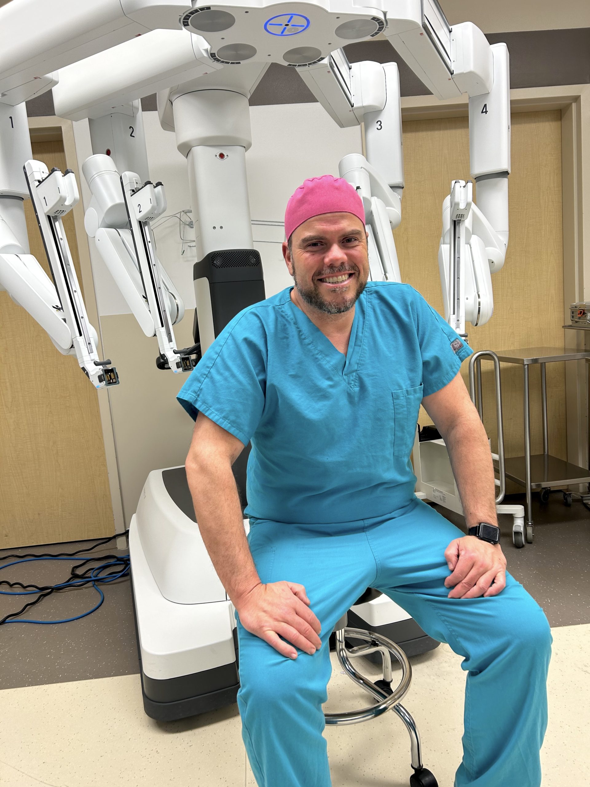
Dr. Romeo Augusto Lucas is a board-certified gynecologist who started the New England Center for Pelvic Health in Freeport, Maine. His clinic focuses on Endometriosis, Pelvic, & Sexual Health. Dr. Lucas wanted to create a place where women with pelvic pain and endometriosis can get complete care. This includes checking everything properly, finding out what’s wrong, and giving the right treatment. His treatments use different methods like hormone therapy, pain relief, nerve treatments, surgery, and hands-on therapy to help with pelvic pain.
If you’re dealing with these health issues, reach out to us. We’re here to help you feel better. Contact us today to learn more.

Now Licensed In Maine,
New Hampshire & Vermont

MINIMALLY INVASIVE SURGERY
Minimally invasive surgeries for Endometriosis, Pelvic, & Sexual Health, performed using Robotic Laparoscopy, result in less pain, rapid recovery, and the best chance for long-lasting results.
Patient Success Stories: Healing with Dr. Lucas
Explore Expert Endometriosis, Pelvic, & Sexual Health
Ready to take the first step towards personalized Endometriosis, Pelvic & Sexual Health care? Fill out our simple form below to schedule a consultation with Dr. Romeo Lucas, and embark on your journey to relief and well-being.
Schedule Appointment Today
Surgery Book Appt
Appointment booking form.
